Explore the World through Microscopes

The RTNN will return to the Museum of Life and Science in Durham, NC on Saturday, August 17th from 10 am – 4 pm.

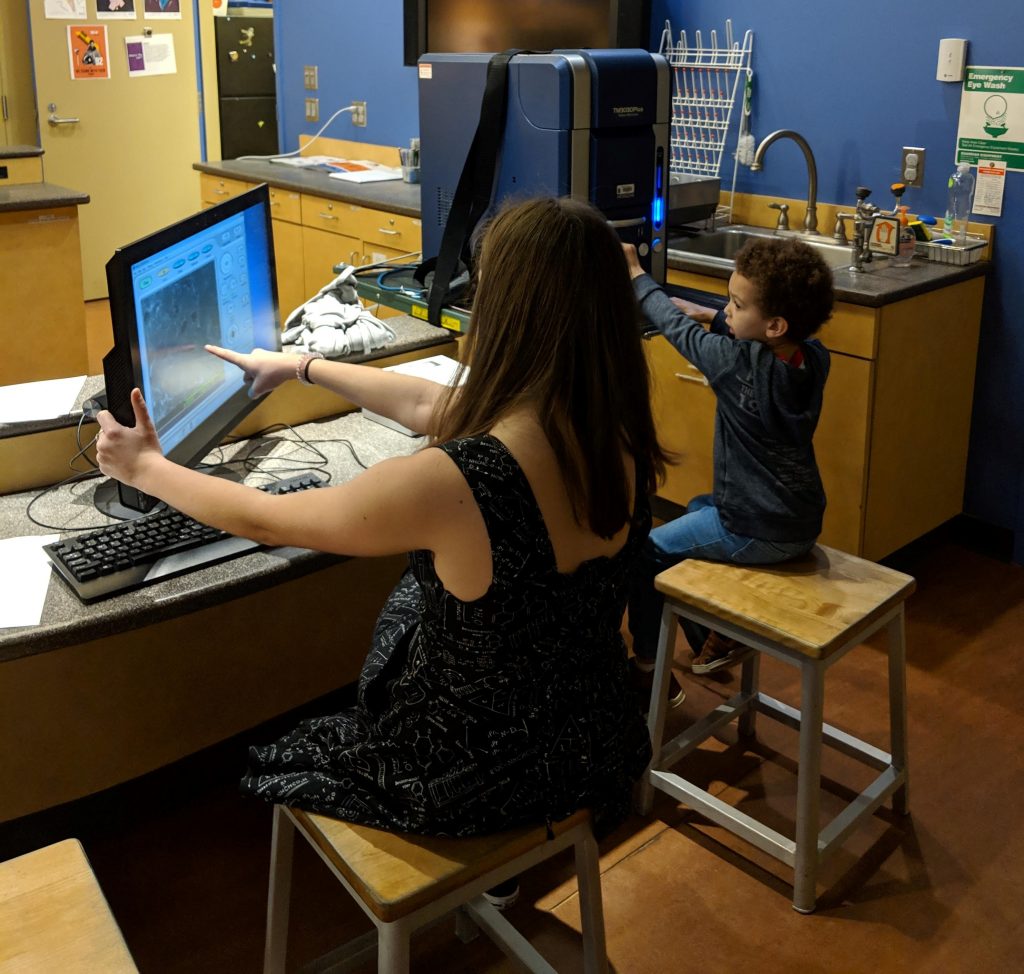
We will investigate things you encounter every day through a different lens! Join us as we take an up-close look at everything from bugs to Band-Aids through a Scanning Electron Microscope! Can you guess what you’re viewing from structures nanometers small?

Learn more about our previous event at the museum here.

A Giant Tool to See the Tiniest Things

The cryo-TEM housed in Duke’s Shared Materials Instrumentation Facility was recently featured in Duke’s Research News. This instrument helps researchers resolve the complex structures of proteins. The cryo-TEM can capture hundreds of thousands of images of these small molecules, and power software is employed to reconstruct the structures in three-dimensions. To learn more, read the full news article.

Museum Patrons Learn the Joys of Microscopy

To celebrate Nanomonth at the Museum of Life and Science, the RTNN brought its expertise as well as a set of light microscopes and a portable desktop scanning electron microscope to The Lab. This venue provides a unique space for hands-on science experiences. Museum visitors learned the differences between the two types of microscopes and examined a variety of samples on each tool. Over 130 people of all ages stopped by to view electronic components found in their phones and tablets and the structures that give Morpho butterfly wings their brilliant blue color. During the NC Science Festival we will be at the Chapel Hill Public Library on April 13th from 1-3 pm with these microscopes and other nanotechnology hands-on activities.

Scanning Electron Microscopy Short Course

February 8, 2019 @ 9:00 am 5:00 pm

The goal for the day is to learn the basics of scanning electron microscopy (SEM) theory and SEM operation so that you can apply that knowledge to analyzing your own samples and/or understanding what SEM data means. Please note that a short course is designed to teach you how the technique works, the data that can be derived thereof, and what the generated data means. Training is designed to teach you the operational specifics of a particular instrument. Assuming that all is well you should be able to drive the Hitachi S-3200N SEM more or less independently by the end of the day. That said, it is not unusual for users to request additional training for a particular instrument.

We will start with an introduction lecture to SEM after which we will move to the lab. In the first lab, the student will observe a demonstration of SEM operation.  Then each of you will drive the microscope on a standard sample and we will explore changing instrument conditions and the resulting effect on the data. During this phase, we will also learn how to focus and correct astigmatism as well as how to properly set signal gain (contrast) and offset (brightness). After a break for lunch, there will be another lecture where we learn more about electron beam-sample interactions, detectors, how to pick imaging conditions, etc. Then we will go to the lab where you can apply what you have learned by imaging any sample you wish. You are welcome to bring a sample for this time or I can find one that is appropriate.

You should bring something to write with and on, a memory stick for the presentation and any data or other information that is electronic, and last but certainly not least, your brain. You can also bring a sample for hands-on time in the afternoon. We will take a short break for lunch, probably on the order of 30-45 minutes, so it might be smart to bring lunch with you.

This should be a relaxed and enjoyable day where you learn something fun and useful. Questions? Please contact Chuck Mooney at cbmooney@ncsu.edu. To maximize hands-on time, the class is limited to three students.

Registration costs: $50 for academic, government, and non-profit participants; $350 for industry participants

To register, add yourself to the short course through AIF’s lab management software, Mendix. Click here to watch a short video on how to register for a short course in Mendix. Select “Sign up for a Short Course.”
2410 Campus Shore Drive
Raleigh, NC United States
+ Google Map

RTNN users win awards at Microscopy & Microanalysis meeting

RTNN congratulates Houston Dycus, Dr. Jim LeBeau, and Matt Cabral for their recent awards at the 2016 Microscopy & Microanalysis meeting. Dycus (left picture), LeBeau, and Cabral (right picture) are all in the Department of Materials Science and Engineering at NC State. Dycus and LeBeau were awarded the best paper in the Microscopy and Microanalysis Journal (Materials Science Category) for their paper: Accurate nanoscale crystallography in real-space using scanning transmission electron microscopy.” Dycus also received the best poster award for his work, “Resolving Atomic Scale Chemistry and Structure at NO and Ba Passivated SiC/SiO2 Interfaces.” Cabral won a Student Scholar award for his contribution entitled: Direct Observation 
of Local Chemistry and Local Cation Displacements in the Relaxor Ferroelectric PMN-PT.”